All About Toxic Mercury Amalgam

The FDA Continues to Fail Our Children
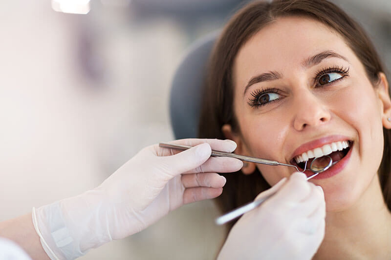
[vc_row][vc_column][us_image image="6297" align="right"][vc_column_text]Why does this government agency whose job is to protect all the citizens of this county, continue to ignore the dangers of mercury amalgam – Especially in our children? This article is from Dr....

How to Protect Yourself from Mercury Amalgam — Even Though the Government and Your Insurance Company Don’t Want You To
[vc_row][vc_column][us_image image="6296" align="right"][vc_column_text]There are powerful forces supporting the continued use of this toxic poison. Here's an article about what you can to oppose these forces. This article is from Dr. Mercola's website. Insurance and...
The Dangerous Health Effects of Fluoride and Mercury Fillings
This video and the following article can be found at Dr. Mercola's website. For Optimal Health, Mind Your Oral Microbiome and Avoid Fluoride, Harsh Mouth Rinses and Amalgam Fillings While often overlooked, your dental health can have a significant impact on...

Petition to Ban Mercury Dental Fillings in Children
Starting in 2018, the European Union has banned the use of mercury amalgam fillings in children under age 15. It is well past time for America to do the same. Here is a call from Consumers for Dental Choice to sign a petition asking the FDA to ban amalgam use in...

Toxic Mercury Release Increases from Wi-Fi Radiation
Previous studies have shown that electromagnetic radiation from MRI's and X-ray exposure increase the release of mercury from amalgam filled teeth. This is the first study to confirm that this also occures with exposure to Wi-Fi devices. Here is a reprint of the...

Mercury Fillings May Cause Breast Cancer
Dr. Veronique Desaulniers - Greenmedinfo.com Thursday, July 12, 2012 8:01 CDT There is no safe level of mercury. Mercury is the most toxic naturally occurring substance on the planet, yet, according to the EPA, there is currently over 1 ton of mercury from amalgam...

Just 1 Single Drop of This Would Poison a Lake Enough to Ban Fishing on It
By Dr. Mercola To see the interview with Charlie Brown I am excited to have the opportunity to interview Charlie Brown, one of my legal heroes in the battle against mercury. Charlie has been a major force in the ground roots movement against mercury amalgams....

The Smoking Time Bomb Sitting Next to Your Brain
Has Its Moment Finally Arrived? By Dr. Mercola - January 28, 2011 At the end of the two-day hearing to evaluate the safety of amalgam, the FDA's own scientific panel - including neurologists, toxicologists, epidemiologists, and environmental health specialists - told...

Are you tired?
An Unspoken Connection: The Link Between Mercury Based Fillings and Chronic Fatigue Are you tired? Have you ever been tired? Have you ever been more than tired, exhausted, totally spent? And I don't mean the kind of tired you get from a hard day of work, a long...

Special Report!
If you have decided to have your toxic mercury fillings removed and you want to protect yourself against the dangers of toxic mercury... BEFORE YOU DO You must read this Special Report: The Hidden Dangers of Amalgam Removal to download Click Here In this report you...